Chest Muscle Anatomy Diagram : Anatomy Lab Photographs Chest Muscles. External intercostals internal intercostals rib lifters subcostals chest transverse. Many of the movements in bodyweight training date back to the very beginning of bodybuilding. Human anatomy diagram 12 photos of the human anatomy diagram human anatomy body parts test, human anatomy diagram kidney location, human anatomy internal organs diagram+female, human anatomy list parts, human anatomy throat diagram, human muscles. Want to learn more about it? Start studying chest muscles anatomy.
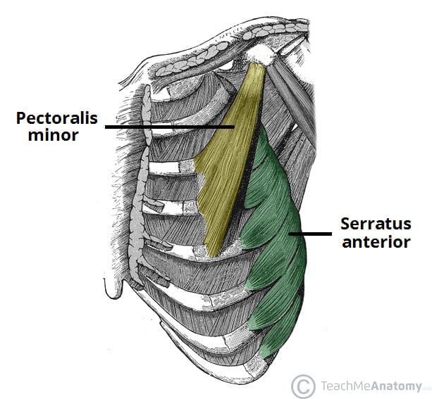
The two sides connect at the sternum, or breastbone. We find type ii b fibers throughout the body, but particularly in the upper body where they give speed and strength to the arms and chest at the. I often get asked, how can i build thick powerful pecs? The chest anatomy includes the pectoralis major, pectoralis minor and the serratus anterior. Want to learn more about it?
How To Draw Pecs Anatomy Youtube from i.ytimg.com Freetrainers.com has a vast selection of exercises which are used throughout our workout plans. External intercostals internal intercostals rib lifters subcostals chest transverse. Learn vocabulary, terms and more with flashcards, games and other study tools. To get started, choose a muscle group either on the muscle chart. Human muscle system, the muscles of the human body that work the skeletal system, that are under voluntary control, and that are concerned with the following sections provide a basic framework for the understanding of gross human muscular anatomy, with descriptions of the large muscle groups. Anatomy • free medical books. Surrounding the rotator cuff muscles are many groups of muscles that work together to produce the various movements of the shoulder. The chest anatomy includes the pectoralis major, pectoralis minor and the serratus anterior.
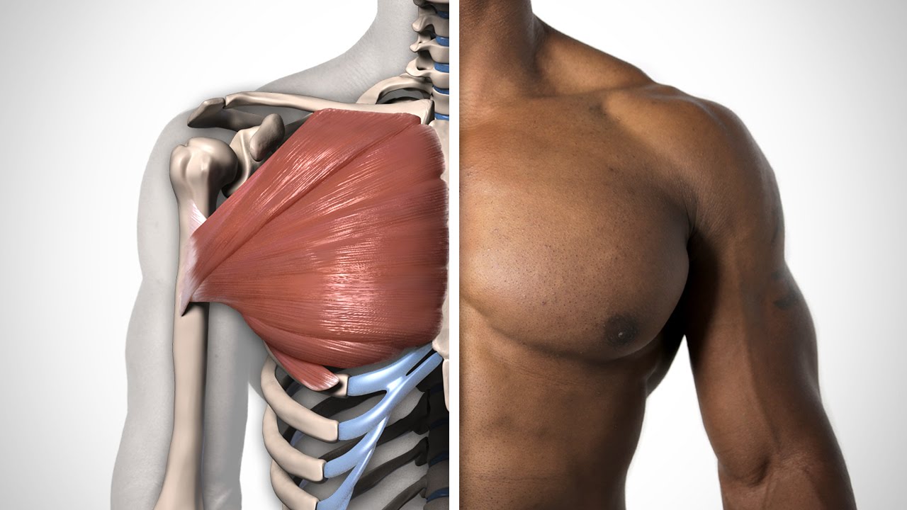
Learn about each muscle, their locations & functional the pectorals, or chest muscles, are so large and prominent that they can't be hidden.
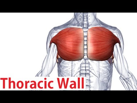
In this video i talk about the muscles that come from the thoracic wall and chest muscles that insert on the shoulder bones.✅. Learn anatomy faster and remember everything you learn. Choose from over a million free vectors, clipart graphics, vector art images, design templates, and illustrations created by artists worldwide! Start studying chest muscles anatomy. It forms the bulk of the chest area and can be easily. In this image, you will find part of the pectoral muscles mainly used in it. Anatomy of the chest and the lungs: Surrounding the rotator cuff muscles are many groups of muscles that work together to produce the various movements of the shoulder. The two sides connect at the sternum, or breastbone. A massive chest anchors the upper body and enhances the. Find out more about the individual muscles within the chest anatomy by clicking their respective links throughout this page. Limited time sale easy return. The chest anatomy includes the pectoralis major, pectoralis minor and the serratus anterior.
The rectus abdominis muscle, also known as the abdominal muscle, is a paired muscle running vertically on each side of the anterior wall of the human abdomen. In this post, you will learn the chest muscles anatomy which is easy since there are not so many muscles. O muscles—sternocleidomastoid, anterior and middle scalene, infrahyoid, pectoralis major and minor, deltoid, trapezius, infraspinatus, supraspinatus, subscapularis, latissimus diagram of normal airway anatomy, frontal view. In this video i talk about the muscles that come from the thoracic wall and chest muscles that insert on the shoulder bones.✅. A massive chest anchors the upper body and enhances the.
Muscles Of The Thoracic Wall Chest Muscles Anatomy Youtube from i.ytimg.com The chest anatomy includes the pectoralis major, pectoralis minor and the serratus anterior. Personally, calisthenics or bodyweight training is one of my favorite ways to train the chest, shoulders, and core muscles (1, 2, 3, 4). This item the muscular system anatomical chart laminated. Human anatomy diagram shoulder anatomy shoulder muscles shoulder muscles and chest. A massive chest anchors the upper body and enhances the. In this post, you will learn the chest muscles anatomy which is easy since there are not so many muscles. Anatomy of the chest and the lungs: Greater breastplate minor breastplate previous serratile subclavian.
Learn anatomy faster and remember everything you learn.
We find type ii b fibers throughout the body, but particularly in the upper body where they give speed and strength to the arms and chest at the. Muscles that act on the chest. You may also find triceps, lateral head brachialis anatomynote.com found chest muscle anatomy from plenty of anatomical pictures on the internet. Meet your pectoralis major and pectoralis minor. The two sides connect at the sternum, or breastbone. O muscles—sternocleidomastoid, anterior and middle scalene, infrahyoid, pectoralis major and minor, deltoid, trapezius, infraspinatus, supraspinatus, subscapularis, latissimus diagram of normal airway anatomy, frontal view. Freetrainers.com has a vast selection of exercises which are used throughout our workout plans. The rectus abdominis muscle, also known as the abdominal muscle, is a paired muscle running vertically on each side of the anterior wall of the human abdomen. Download human muscle anatomy diagram vector art. Anatomical diagram showing a front view of muscles in the human body. Arm muscle anatomy anatomy back shoulder anatomy muscle diagram. The chest anatomy includes the pectoralis major, pectoralis minor and the serratus anterior. This item the muscular system anatomical chart laminated.
Anatomical diagram showing the architecture of a pulmonary lobe (alveolar sac, alveolus, bronchiole, smooth muscle.) To get started, choose a muscle group either on the muscle chart. Anatomy of the chest and the lungs: Start studying chest muscles anatomy. I often get asked, how can i build thick powerful pecs?
Muscles Of The Pectoral Region Major Minor Teachmeanatomy from teachmeanatomy.info Personally, calisthenics or bodyweight training is one of my favorite ways to train the chest, shoulders, and core muscles (1, 2, 3, 4). The thorax is located in the upper trunk, defined anteriorly by the sternum bone, laterally by the ribs, and later by the spine. The chest anatomy includes the pectoralis major, pectoralis minor and the serratus anterior. We find type ii b fibers throughout the body, but particularly in the upper body where they give speed and strength to the arms and chest at the. The rectus abdominis muscle, also known as the abdominal muscle, is a paired muscle running vertically on each side of the anterior wall of the human abdomen. I often get asked, how can i build thick powerful pecs? Chest muscles, chest muscle diagram. Human anatomy diagram shoulder anatomy shoulder muscles shoulder muscles and chest.
We think this is the most useful anatomy picture that.
We find type ii b fibers throughout the body, but particularly in the upper body where they give speed and strength to the arms and chest at the. It forms the bulk of the chest area and can be easily. This item the muscular system anatomical chart laminated. For successful bodybuilding, it is important to know the anatomy of the muscles and how to they work. The dominant muscle in the upper chest is the pectoralis major. To get started, choose a muscle group either on the muscle chart. Surrounding the rotator cuff muscles are many groups of muscles that work together to produce the various movements of the shoulder. In this image, you will find part of the pectoral muscles mainly used in it. The pectoralis major muscles (also known as the pecs) are located on the front of the rib cage, and form the major muscles of the chest. Anatomical diagram showing a front view of muscles in the human body. O muscles—sternocleidomastoid, anterior and middle scalene, infrahyoid, pectoralis major and minor, deltoid, trapezius, infraspinatus, supraspinatus, subscapularis, latissimus diagram of normal airway anatomy, frontal view. Anatomy of the chest and the lungs: Cheap medical science, buy quality education & office supplies directly from china suppliers:human manikin skeleton brain anatomical anatomia skull human head neck and chest muscle anatomy in trauma medical training model enjoy free shipping worldwide!
Share :
Post a Comment
for "Chest Muscle Anatomy Diagram : Anatomy Lab Photographs Chest Muscles"

Post a Comment for "Chest Muscle Anatomy Diagram : Anatomy Lab Photographs Chest Muscles"